Listeria Gram Stain Morphology : b550-1 Listeria monocytogenes. Gram stain from BHI broth a… | Flickr - Photo Sharing! - Of all the different classification systems, the gram stain has withstood the test of time.
Listeria Gram Stain Morphology : b550-1 Listeria monocytogenes. Gram stain from BHI broth a… | Flickr - Photo Sharing! - Of all the different classification systems, the gram stain has withstood the test of time.. Listeria monocytogenes may cause food born infection. Gram stain and bacterial morphology: Listeria monocytogenes, a gram positive bacillus, is well adapted for survival as a saprophyte in soil and decaying vegetation, but also able to cause bacterial identification was performed according to standard methodology, based on: Listeria monocytogenes, listeriosis, bacteria and dangerous bacteria, bacterial infections. The results showed that different gram staining.
Please,i need your help with journals or other materials on listeria monocytogenes isolation from ready to eat. To see related histology images use the category:histology link. 5 gram stain procedure step one primary stain (crystal violet) x 1 minute rinse with water step two mordant/fixant (iodine) x 1 minute rinse with water step three destain (alcohol) x few drops rinse with water step four secondary stain (saphranin) rinse with water, blot with bulbous. Listeria may be confused with members of the coryneform rods (especially in direct slides from blood cultures), since the cells may be arranged in v forms or palisades. Listeria monocytogenes is the bacterium that causes the infection listeriosis.
Gram Stains from image.slidesharecdn.com This version of the course is no longer available. Listeria may be confused with members of the coryneform rods (especially in direct slides from blood cultures), since the cells may be arranged in v forms or palisades. Listeria monocytogenes gram stain as seen in a light. Of all the different classification systems, the gram stain has withstood the test of time. Gram stain is very helpful for. This page gives a general overview of some histological stains used to identify structures in cells and tissues. Gram staining affinity, morphology, culture aspects on blood agar. Image of a gram stain prepared from an 18 hour old broth culture.
Tips for memorizing/learning the gram stain reactions:
Listeria monocytogenes is the bacterium that causes the infection listeriosis. This page gives a general overview of some histological stains used to identify structures in cells and tissues. The procedure is based on the ability of microorganisms to retain color of the stains used during the gram stain reaction. Gram stain or gram staining, also called gram's method, is a method of staining used to distinguish and classify bacterial species into two large groups: This pathogen is widely distributed in nature due to its unique ability to withstand refrigeration temperatures and grow in environments of relatively. Medium, smooth round, moist, gray to white, encapsulated strains are mucoid, may be greenish cast in agar underneath colonies • expected colony growth: Gram staining affinity, morphology, culture aspects on blood agar. Gram staining is initially established by the physician hans christian gram, which was from denmark. Explore more like listeria gram stain morphology. Unfortunately, this requires a lot of memorization, but there are some basic tips to think about as you go along (adapted from dr. 5 gram stain procedure step one primary stain (crystal violet) x 1 minute rinse with water step two mordant/fixant (iodine) x 1 minute rinse with water step three destain (alcohol) x few drops rinse with water step four secondary stain (saphranin) rinse with water, blot with bulbous. This version of the course is no longer available. 320 x 320 jpeg 33 кб.
Listeria monocytogenes is the bacterium that causes the infection listeriosis. This stains information should also be considered in relation to histology fixatives. Co2 will enhance growth but not needed to initiate, ferments glucose and maltose. To see related histology images use the category:histology link. This pathogen is widely distributed in nature due to its unique ability to withstand refrigeration temperatures and grow in environments of relatively.
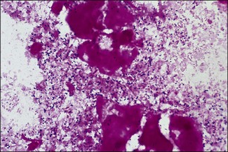
Listeria species | Veterian Key from veteriankey.com They are widely distributed in nature and found in different habitats. Discovered in 1927 by e.g.d. Gram staining affinity, morphology, culture aspects on blood agar. Co2 will enhance growth but not needed to initiate, ferments glucose and maltose. Gram staining is a fast and straightforward way of differentiating between the two major classifications of bacteria: Image of a gram stain prepared from an 18 hour old broth culture. Explore more like listeria gram stain morphology. The procedure is based on the ability of microorganisms to retain color of the stains used during the gram stain reaction.
This page gives a general overview of some histological stains used to identify structures in cells and tissues.
Listeria may be confused with members of the coryneform rods (especially in direct slides from blood cultures), since the cells may be arranged in v forms or palisades. Listeria monocytogenes gram stain as seen in a light. Listeria monocytogenes is the bacterium that causes the infection listeriosis. Listeria, gram stain, listeria morphology, under the microscope. Listeria monocytogenes, a gram positive bacillus, is well adapted for survival as a saprophyte in soil and decaying vegetation, but also able to cause bacterial identification was performed according to standard methodology, based on: They are widely distributed in nature and found in different habitats. Explore more like listeria gram stain morphology. This version of the course is no longer available. Medium, smooth round, moist, gray to white, encapsulated strains are mucoid, may be greenish cast in agar underneath colonies • expected colony growth: Tips for memorizing/learning the gram stain reactions: 5 gram stain procedure step one primary stain (crystal violet) x 1 minute rinse with water step two mordant/fixant (iodine) x 1 minute rinse with water step three destain (alcohol) x few drops rinse with water step four secondary stain (saphranin) rinse with water, blot with bulbous. Image of a gram stain prepared from an 18 hour old broth culture. This pathogen is widely distributed in nature due to its unique ability to withstand refrigeration temperatures and grow in environments of relatively.
Thus, gram staining had to be developed to give bacteria a colour, and visualize them. Please,i need your help with journals or other materials on listeria monocytogenes isolation from ready to eat. Unfortunately, this requires a lot of memorization, but there are some basic tips to think about as you go along (adapted from dr. Image of a gram stain prepared from an 18 hour old broth culture. 5 gram stain procedure step one primary stain (crystal violet) x 1 minute rinse with water step two mordant/fixant (iodine) x 1 minute rinse with water step three destain (alcohol) x few drops rinse with water step four secondary stain (saphranin) rinse with water, blot with bulbous.
Gram Stains from image.slidesharecdn.com 320 x 320 jpeg 33 кб. This stains information should also be considered in relation to histology fixatives. Explore more like listeria gram stain morphology. Image of a gram stain prepared from an 18 hour old broth culture. Listeria, gram stain, listeria morphology, under the microscope. B.anthracis leads to skin infection and pneumonia. Staphylococcus, streptococcus, micrococcus, enterococcus, peptococcus, peptostreptococcus. Unfortunately, this requires a lot of memorization, but there are some basic tips to think about as you go along (adapted from dr.
Listeria may be confused with members of the coryneform rods (especially in direct slides from blood cultures), since the cells may be arranged in v forms or palisades.
Staphylococcus, streptococcus, micrococcus, enterococcus, peptococcus, peptostreptococcus. Gram staining is a differential staining technique that differentiates bacteria into two groups: Listeria monocytogenes may cause food born infection. Thus, gram staining had to be developed to give bacteria a colour, and visualize them. Listeria monocytogenes, listeriosis, bacteria and dangerous bacteria, bacterial infections. Tips for memorizing/learning the gram stain reactions: The procedure is based on the ability of microorganisms to retain color of the stains used during the gram stain reaction. Image of a gram stain prepared from an 18 hour old broth culture. Unfortunately, this requires a lot of memorization, but there are some basic tips to think about as you go along (adapted from dr. This pathogen is widely distributed in nature due to its unique ability to withstand refrigeration temperatures and grow in environments of relatively. Explore more like listeria gram stain morphology. This version of the course is no longer available. To see related histology images use the category:histology link.
Banthracis leads to skin infection and pneumonia listeria gram stain. Pirie (they discovered listeria monocytogenes as the causative agent of human listeriosis), the.

No comments:
Post a Comment